
Myositis associated with connective tissue disease:
The classic ones – Antisynthetase Syndrome
The less well studies ones – Scleromyositis
Werner Stenzel
Consultant, lecturer of Neuropathology at the Department of Neuropathology, Charité – Universitätsmedizin, Berlin, Germany
Head of the Neuromuscular Diagnostic and Research Unit at the Department of Neuropathology, Charité – Universitätsmedizin, Berlin, Germany
Head of educational program for medical students at the Department of Neuropathology
In this seminar, the so-called overlap myositis will be highlighted.
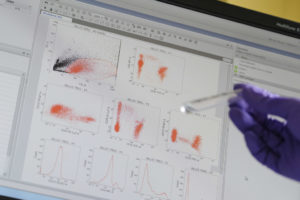
I will first address, anti-synthestase syndromes as a classical example of this group where a typical form of myositis is associated with characteristic skin lesions and interstitial lung disease. This group is an entity on its own and can well be characterized based on the additional typical presence of t-RNA-synthetase autoantibodies like anti-Jo1. We have recently identified presence of plasma cells in the skeletal muscle tissue of these patients. Here I will present our recent results showing a highly specialized immunological niche as extra medullary immunological niche for plasma cells and activated B cells. It contains alkaline phosphatase-positive perimysial fibroblasts, CD138+ plasma cells and CXCL12+ and CXCL13+CD20+ B cells. We localize these cells close to CD68+MHC cl. II+CD169+ macrophages and define the MHC cl. I+ and MHC cl. II+ MxA negative type II interferon driven milieu of myofiber activation. Overall, we provide a conceptual framework for antibody producing cells residing in an extramedullary niche and self-perpetuating an autoimmune process in the skeletal muscle.
Second, I will focus briefly on Systemic sclerosis, a chronic disease of connective tissues characterized by fibosis, vasculopathy and autoimmunity. Affected patients show signs of the skin, internal organs and sometimes overlap myositis. The vasculopathy is considered obliterative but the pathogenic basis is not known to date.
We have applied a new electron microscopical technique called large scale electron microscopy allowing a ‘pan and zoom’ approach similar to ‘google earth’ viewing.
This analysis allows to study >1000 capillaries of patients and controls, highlighting reduplications of basement membranes endothelial activation and pericytes proliferations
We show that this type of ultrastructural changes is specific for a subtype of scleromyositis and discus possible pathogenic mechanisms.