A venir
No events are found.
Archives
Participer à la réunion Zoom
https://zoom.us/j/96120152633?pwd=TvOSNd2wci6gQUtstj4gx3FbQ75bRa.1
ID de réunion: 961 2015 2633
Code secret: 393712
« Mitochondrial diseases: pathogenesis and cure »
Dr Metodi METODIEV (Invited by Marc Bitoun)
Institut Imagine, Paris
I am a Chargé de Recherche at INSERM, based at the Imagine Institute for Genetic Diseases in Paris. My training is in mitochondrial biology and mouse genetics, and my work centers on the mechanisms that drive mitochondrial disease. In particular, I study how defects in mitochondrial gene expression and proteostasis disrupt organelle function and lead to pathology, using a combination of cellular systems and animal models. More recently, my research has expanded to translational efforts, including the development of gene-replacement therapy for mitochondrial disorders.

Format hybride Amphi Charcot et zoom
Participer à la réunion Zoom
https://zoom.us/j/99265721001?pwd=ExeaDj7EFGBWVh2fOeur1mljbGWaGT.1
ID de réunion: 992 6572 1001
Code secret: 492332
« Fast subtype myofiber specialization: motoneuron-myofiber crosstalk »
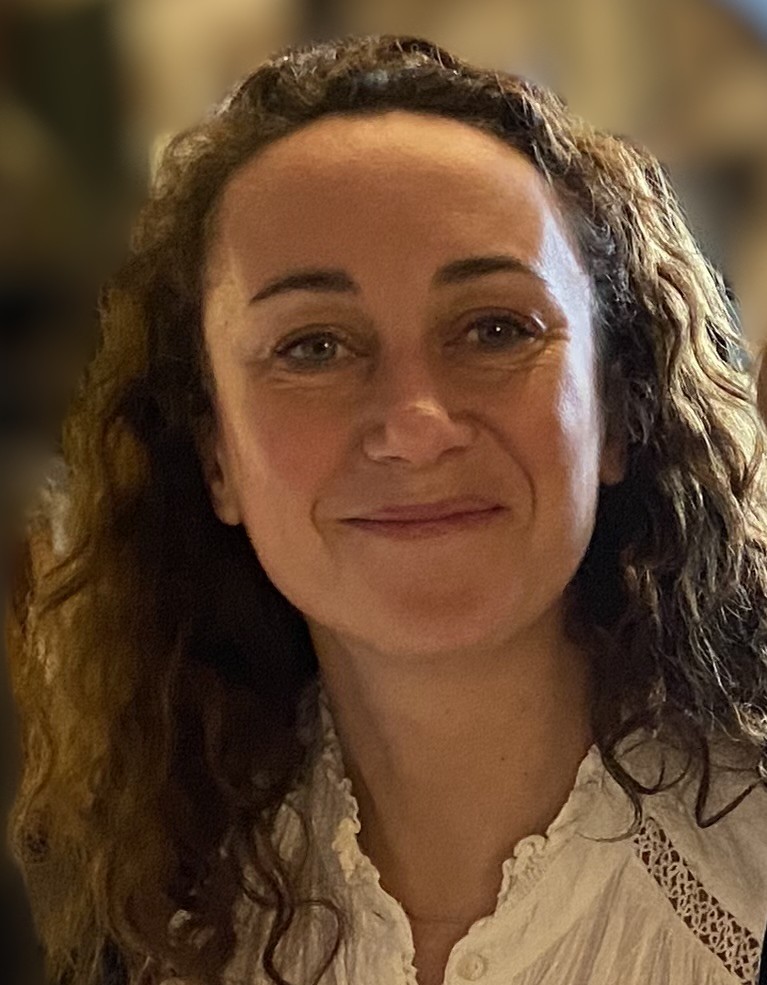
Dr Pascal MAIRE
Institut Cochin. INSERM U1016, CNRS UMR 8104, U.Paris Cité.
Physiopathology of the neuromuscular system
https://zoom.us/j/95857382584?pwd=CGgEbVbUk7JE8TuoVQMxiGZObbzglo.1
ID de réunion: 958 5738 2584
Code secret: 102480
Timing Matters: how the macrophage Clock regulates Skeletal Muscle Regeneration
Yasmine Sebti, PhD, HDR
Inserm UMR1011 & UDSL, Université Lille Nord de France

Participer à la réunion Zoom
https://zoom.us/j/99840003505?pwd=EECZ6aUdnvNGVDazrbHIJbmYHlCyFz.1
ID de réunion: 998 4000 3505
Code secret: 675427
The seminar will be in French with slides in English
HYBRIDE version in Amphi CHARCOT (Pitié Salpêtrière Hôpital) and on zoom
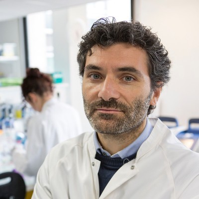
Circulating free DNA and diagnosis – Focus on non-invasive prenatal diagnosis of monogenic diseases
Dr Juliette NECTOUX
Invited by Gisèle Bonne and Tanya Stojkovic
https://zoom.us/j/99748527301?pwd=0QIcHIUSLvc9l9MowegpnAFpsoabqg.1
ID de réunion: 997 4852 7301
Code secret: 959196
Gene Editing for DMD Treatment
Dr Mario AMENDOLA
DR2 Inserm à Genethon, dirige un groupe travaillant au développement d’outils innovants de transfert génique et d’édition du génome, ainsi qu’à leurs applications pour le traitement des maladies génétiques.
https://www.genethon.fr/notre-science/nos-equipes/equipe-edition-du-genome/
ONLY VIRTUAL ON ZOOM
https://zoom.us/j/97539616357?pwd=NiLLYqOGCbOZfGYoJJoEiJZIUI6TJo.1
ID de réunion: 975 3961 6357
Code secret: 621045
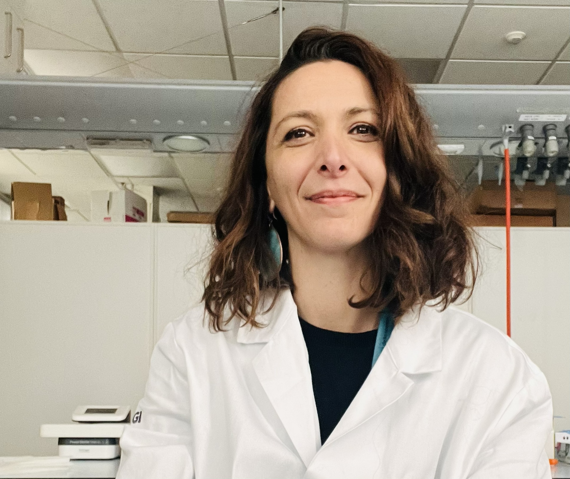
Advanced organoids for studying the human neuromuscular system in vitro
Dr Anna URCIUOLO
ONLY VIRTUAL ON ZOOM
Biosketch: Urciuolo_IM_Seminar_2025
Zoom link => https://zoom.us/j/91502840242?pwd=L6ElWL2j2T7aI5an7RVkUmabzVSEUb.1
AAV.U7snRNA as a platform to treat neuromuscular disorders
Dr Nicolas Wein
https://www.univ-nantes.fr/nicolas-wein
Zoom link => https://zoom.us/j/98751708455?pwd=D31Fj6KO9J2Do2LKskqBDHQBlTCbbB.1
ID de réunion: 987 5170 8455 Code secret: 257035
Skeletal muscle as an endocrine organ during exercise.
Dr Cedric Moro
Cedric Moro is currently Research Director at Inserm, and head of the Team MetaDiab “Pathophysiology of Metabolic Disorders and Diabesity” at the Institute of Metabolic and Cardiovascular Diseases in Toulouse. Our team uses a highly translational medicine approach of the study of metabolism combining innovative cell and mouse models to decipher molecular mechanisms of metabolic disorders, and clinical studies in nutrition and exercise with deep phenotypic investigation. The team aims at understanding the biological determinants and molecular mechanisms of metabolic disorders in various pathophysiological contexts (obesity, aging, type 2 diabetes, physical inactivity). Using a highly integrative bedside-to-bench approach, we investigate novel targets and mechanisms in cell and mouse models as well as in humans. Our projects are focused on the underlying mechanisms of insulin resistance, lipid droplet and metabolic dysfunction in skeletal muscle and adipose tissue, as well as their crosstalk in metabolic regulation and diseases.
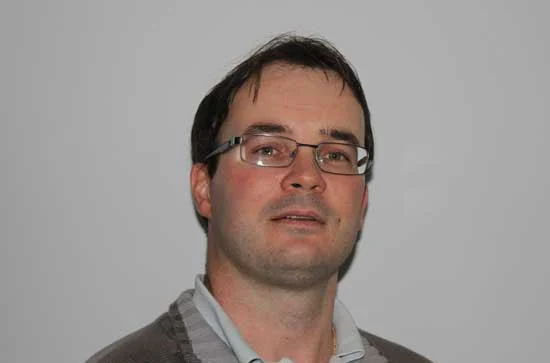
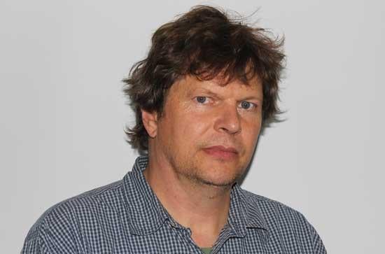
Title: Proteomic of muscle aging (Kay Ohlendieck) and Proteomics: past, present and future (Paul Dowling)
du Department of Biology, Maynooth University, National University of Ireland

Zoom link => https://zoom.us/j/94208295145
