A venir
No events are found.
Archives
Early biomarkers of muscle atrophy and of neuromuscular maladaptation during short-term inactivity in humans
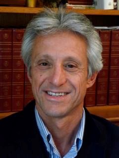
Marco Narici
Professor at the University of Padova, Italy.
Marco Narici (PhD) is Professor in Physiology at the University of Padova, Italy. He worked as researcher at the Rodolfo Margaria Laboratory, University of Milan, Italy, and took up the position of Assistant Professor at the Medical Research Centre of Geneva University, CH (1994-96). In 1999 he moved back to the UK as Professor in Physiology of Ageing at Manchester Metropolitan University where he was Director of the Institute of Research into Human Movement. In 2012, he was appointed as Professor in Clinical at the University of Nottingham to work at the MRC ARUK Centre for Musculoskeletal Ageing. In 2017 he moved back to Italy, to the University of Padova where he took the chair in Physiology at the School of Medicine. He has been coordinator of the EC FP5 project BETTER AGEING and leader of two WPs of FP7 Project MYOAGE, he has been part of EC Framework 7 Advisory Panels for Ageing Research (LinkAge, WhyWeAge) and was President of the European College of Sport Science (2013-15). He has published over 230 peer reviewed journal articles (H-factor 64) and book chapters. His present work and interests are focused on the mechanisms of remodeling of human neuromuscular system with exercise, inactivity (including spaceflight) and ageing. He is presently coordinating the NeuAge PRIN Project funded by the Italian Ministry of Education, University and Research (MIUR) focusing on the Mechanisms of Neuromuscular Ageing and its Functional Implications.
Read more information on Marco Narici’s research group.
Register by email.
« Ex vivo editing of human hematopoietic stem cells for erythroid expression of therapeutic proteins”
Mario Amendola
Chargé de recherche INSERM, Responsable de l’équipe » genome editing’ group, UMR_S951, Evry, FR.
Targeted genome editing has a great therapeutic potential to treat disorders that require protein replacement therapy. To develop a platform independent of specific patient mutations, therapeutic transgenes can be inserted in a safe and highly transcribed locus to maximize protein expression. Therefore, we developed an ex vivo editing approach to achieve efficient gene targeting in human hematopoietic stem/progenitor cells (HSPCs) and robust expression of clinically relevant proteins by the erythroid lineage. Using CRISPR-Cas9, we integrated different transgenes under the transcriptional control of the endogenous α-globin promoter, recapitulating its high and erythroid-specific expression. Erythroblasts derived from targeted HSPCs secrete different therapeutic proteins, which retain enzymatic activity and cross-correct patients’ cells. Moreover, modified HSPCs maintain long-term repopulation and multilineage differentiation potential in transplanted mice. Finally, additional optimization of therapeutic transgene, CRISPR-Cas9 tools and donor DNA delivery further improve efficiency and safety of the approach.
Overall, we establish a safe and versatile CRISPR-Cas9-based HSPC platform for different therapeutic applications, including hemophilia and inherited metabolic disorders.
You can find more information on Mario Amendola on his Biosketech.
Role of skeletal muscle mesenchymal progenitors in bone regeneration
Céline Colnot
Directrice de Recherche DR2 INSERM à l’Institut Henri Mondor de Recherche Biomédicale
Skeletal muscle and bone exhibit great capacities to regenerate due to tissue-specific stem cells, i.e. satellite cells and skeletal stem cells from periosteum and bone marrow. However, bone fails to heal properly in 10% of bone injuries and delayed healing is increased to 40% in patients with soft tissue damage associated with bone fracture. The role of skeletal muscle in bone repair is well recognized clinically but the underlying cellular and molecular mechanisms are poorly understood. Muscle regulates the inflammatory environment of fracture and muscle satellite cells are providing a source of growth factors for bone repair.
We show that mesenchymal progenitors residing in skeletal muscle adjacent to bone also participate in cartilage and bone formation during bone regeneration. We used single cell RNAseq analyses to characterize the skeletal muscle mesenchymal progenitors, their response to bone injury and the impact of musculoskeletal trauma on the cellular response to injury. The results elucidate the role skeletal muscle during bone repair as a source of mesenchymal progenitors contributing to bone repair, and as a mediator of initial fibrotic response and fibrotic remodelling in bone repair. The findings suggest that new pharmacological and cell-based approaches can be developed to improve musculoskeletal regeneration by targeting skeletal muscle adjacent to bone.
Read more information on Céline Colnot’s research group and on her biosktech.

Register by email.
« The role of SMN outside the CNS: Implications for clinical care of Spinal Muscular Atrophy”
Rashmi Kothary
Deputy Scientific Director, Ottawa Hospital Research Institute, Professor, Departments of Medicine, CMM, and BMI, University of Ottawa
You can find more information on Rashmi Khotary on his Biosketech and on his website.

Epigenetic control of the satellite cell response to muscle injury
Jeffrey Dilworth
Senior Scientist, Ottawa Hospital Research Institute
Summary
In response to injury, Muscle Stem Cells (MuSCs) exit quiescence, proliferate and differentiate to form new myofibers, while also repopulating the stem cell niche in preparation for future injuries. The transitioning between cell fates that allow MuSCs to regenerate the muscle fibers involves temporal ordered changes in gene expression that occur in response to environmental cues. The environmental cues are interpreted by the epigenetic machinery in the cell to establish transcriptional permissive or transcriptional repressive states at individual genes. The Dilworth lab is working to understand the role for different epigenetic enzymes in regulating the transitions between MuSC cell fates. In this seminar, characterization of the role for the H3K27me3 demethylase JMJD3 and the transcription elongation factor NELF in regulating MuSC-mediated regeneration will be presented.
Read more on Jeffrey Dilworth’s biosketch
Register by email.
Integrative and single-cell genomics approaches to dissect cell fate decisions and genetic disorders
Davide Cacchiarelli
Leader of the Laboratory of Integrative Genomics at TIGEM (Telethon Institute of Genetics and Medicine), Assistant Investigator, Assistant Professor of Molecular Biology, Department of Translational Medicine, University of Naples Federico II, Italy
Our lab aims to identify the mechanisms controlling cell fate decisions during cellular differentiation, conversion and reprogramming, and how such processes are affected by genetic mutations of key regulatory proteins including transcription factors. To achieve this goal he proposes to integrate descriptive, functional and single-cell genomics to dissect how genetic elements and their variants impinge on the temporal and spatial control of gene expression.
Short CV:
Academics:
- Oct 07 – Jul 11 Microsoft Research PhD student – Lab of Irene Bozzoni
- Oct 11 – Feb 17 Research Fellow (Human Frontier Science Program) – Lab of Alex Meissner and Eric Lander
- Feb 17 – TIGEM Assistant Investigator / Armenise/Harvard Fellow / Rita-Levi Montalcini Assistant Professor of Molecular Biology at the University of Naples « Federico II »
Research Grants:
- 2017 – Rita Levi-Montalcini Assistant Professorship Grant (Tenure-Track)
- 2017 – European Research Council (ERC) Starting Grant
- 2017 – Telethon Foundation Start-up Grant
- 2017 – Armenise/Harvard Career Development Award
Research Topics:
- Single Cell Genomics
- Stem Cell Reprogramming and Transdifferentiation
- High-Throughput dissection of CIS regulatory elements and TFs
More information on his website
Register by email.
Mechanical Tension Drives Cell-Cell Fusion
Elizabeth Chen
Professor of Molecular Biology and Cell Biology, Hamon Center for Regenerative Science and Medicine, University of Texas Southwestern Medical Center
You can find more information on Elizabeth Chen on his Biosketch.
Register by email.
Role of the circadian biological clock in muscle physiology
Hélène Duez
Univ. Lille, Inserm, CHU Lille, Institut Pasteur de Lille, U1011- EGID, F-59000 Lille, France
The biological clock has long been known to play crucial roles in several aspects of physiology, and clock disruption, as seen in shiftwork, frequent jetlag or extended light exposure and feeding period, increases the risk of developing a myriad of pathologies including metabolic and inflammatory disorders. Focusing on the clock component and drug targetable Rev-erbα nuclear receptor, our goal is to unravel the cellular and molecular mechanisms by which the clock impacts metabolism and inflammation in several patho-physiological contexts, particularly muscle diseases.
We reported that Rev-erb-α controls skeletal muscle mitochondrial biogenesis and oxidative function through enhanced Lkb1-Ampk-Sirt1-Pgc1α signaling. As a result, Rev-erbα deficiency led to decreased skeletal muscle mitochondrial content and aberrant morphology of the remaining mitochondrial network with increased autophagy, ultimately leading to compromised exercise capacity. Interestingly, pharmacological Rev-erb-α activation in skeletal muscle in vivo increases skeletal muscle mitochondrial respiration, energy expenditure and exercise capacity, identifying Rev-erb-α as a target to improve muscle function. In parallel, we studied muscle biopsies from healthy or obese/diabetic patients taken ‘around the clock’. Our results show that mitochondrial function oscillates throughout the 24h cycle in human myotubes, and that oscillations in clock genes are impaired when myotubes are isolated from diabetic patients. More recently, we showed that circadian misalignment in healthy young lean men compromises skeletal muscle insulin sensitivity, suggesting that impaired skeletal muscle clock function may contribute to the increased risk of type 2 diabetes observed in shiftworkers.
We also recently showed that Rev-erbα deficiency results in muscle mass loss and reduced fiber size. We found that administration of a Rev-erb agonist in the right time window is able to reverse dexamethasone-induced muscle atrophy, identifying Rev-erb-α as a promising target to limit muscle loss in patients with chronic inflammatory diseases treated with glucocorticoids. It remains to be determined whether Rev-erb-α activation is efficient at limiting muscle wasting in other contexts (eg. obesity/diabetes, cachexia, myopathy).
We are pursuing our research line by studying the role of Rev-erbα, and more generally of the clock, in sarcoplasmic reticulum homeostasis and calcium fluxes on one side, and in muscle regeneration after injury on the other. Our results point to a role of the clock in both muscle stem cells and infiltrating immune cells (ongoing).
Altogether these data identify the clock component Rev-erb-α as an interesting target for the treatment of muscle diseases.
CURRICULUM VITAE Hélène Duez
Hélène Duez : DR2 Inserm, Responsable de l’équipe 5 « Récepteurs nucléaires et rythmes circadiens en physio-pathologie » à l’Institut Pasteur de Lille.
Après une thèse à l’Institut Pasteur de Lille et un post-doctorat à Toronto où j’ai étudié, respectivement, le rôle des récepteurs nucléaires dans le métabolisme des lipides, puis les dérégulations du métabolisme lipidique chez le sujet diabétique, je me suis établie à l’Institut Pasteur de Lille au sein de l’U1011 pour m’intéresser à l’implication de l’horloge biologique dans la physio-pathologie.
On sait depuis longtemps que l’horloge biologique joue un rôle crucial dans plusieurs aspects de la physiologie. La perturbation de l’horloge, telle qu’elle est observée dans le travail posté, les décalages horaires fréquents, l’exposition prolongée à la lumière artificielle le soir/la nuit et la prise alimentaire en dehors de la phase de jour, augmente le risque de développer une myriade de pathologies, notamment des troubles métaboliques (obésité, diabète de type 2, NASH), inflammatoires et cardio-vasculaires (athérosclérose, infarctus du myocarde). En se concentrant sur les Rev-erbs et les ROR, notre objectif est de démêler les mécanismes cellulaires et moléculaires par lesquels l’horloge influe sur le métabolisme et l’inflammation dans plusieurs contextes physiopathologiques, en particulier les maladies métaboliques, cardio-vasculaires et musculaires. Nous cherchons également à déterminer si et comment la modulation pharmacologique de ces récepteurs nucléaires empêche la perturbation de l’horloge ou rétablit la rythmicité circadienne dans les gènes/protéines/médiateurs inflammatoires/métabolites et améliore ces conditions pathologiques.
Synaptic mechanisms of neurodegeneration and neuroprotection in Amyotrophic Lateral Sclerosis
Francesco Roselli
Invitée : Maria-Grazia Biferi
Muscles have feelings too: muscle spindle function in normal and dystrophic muscle
Coordinated movements, including locomotion, and their control, require proprioceptive information, i.e. information about muscle tone as well as position and movement of the extremities in space. Muscle spindles are the primary proprioceptive sensory receptors and are present in almost all skeletal muscles. In many neuromuscular diseases, movement is impaired, although the mechanism remains elusive. For example, in muscular dystrophies (MD), patients often experience sudden spontaneous falls, balance problems, as well as gait and posture abnormalities, suggesting the possibility of an impaired muscle spindle function. To investigate, if proprioception is affected in dystrophic muscles, we analyzed muscle spindle number, morphology and function in wildtype mice and in murine models for two distinct types of muscular dystrophy with very different disease etiology, i.e. dystrophin- (DMDmdx) and dysferlin-deficient mice. The total number and the overall structure of muscle spindles in soleus muscles of the dystrophic mice appeared unchanged, demonstrating that intrafusal fibers are less affected by the degeneration compared to extrafusal fibers. Immunohistochemical analyses of wildtype muscle spindles revealed a concentration of dystrophin and b-dystroglycan in intrafusal fibers outside the region of contact to the sensory neuron. Utrophin was substantially upregulated in dystrophin-deficient mice, suggesting a potential compensatory activity of utrophin in DMDmdx mice. Single-unit extracellular recordings of sensory afferents from muscle spindles of the extensor digitorum longus muscle revealed that muscle spindles from both dystrophic mouse strains have an increased resting discharge and a higher action potential firing rate during sinusoidal vibrations. In contrast, the response to ramp-and-hold stretches appeared mostly unaltered compared to the respective wildtype mice. These results show alterations in muscle spindle afferent responses in dystrophic mouse muscles, which might cause an increased muscle tone, and might contribute to the unstable gait and frequent falls observed in patients with muscular dystrophy.
CURRICULUM VITAE Prof. Dr. STEPHAN KRÖGER
Academic Training
1979 – 1984 Studies of Biochemistry at the University of Tübingen
1984 „Diploma“ in Biochemistry
1984 – 1986 masters work at the Max-Planck-Institute for Developmental Biology, Tübingen
1986 – 1989 PhD thesis at the Max-Planck-Institute for Developmental Biology, Tübingen.
1989 – 1992 Postdoctoral fellow at the Department of Neurobiology, Stanford University School of Medicine.
1992 – 2000 Head of an independent research laboratory at the Max-Planck-Institute for Brain Research, Frankfurt.
2000 – 2006 Group leader at the Institute for Physiological Chemistry at the University of Mainz (Faculty of Medicine).
2006 – 2007 Associate Professor for Molecular Cell Biology at the University of Applied Sciences in Senftenberg
since 2007 Professor for Neurophysiology at the Biomedical Center (Medical School), Ludwig-Maximilians-University, Munich
Areas of Research Interests:
Molecular and cellular basis of synapse formation; pathological changes at the neuromuscular junction and in the developing CNS; role of the synapse organizer agrin at the neuromuscular junction and in the CNS; muscle spindle structure, function and development in wildtype and dystrophic mice
Selected recent Publications:
Gerwin, L., Rossmanith, S., Haupt, C., Schultheiß, J., Brinkmeier, H., Bittner, R.E., Kröger, S. (2020) Impaired Muscle Spindle Function in Murine Models of Muscular Dystrophy. J. Physiol. In press
Gerwin, L., Haupt, C., Wilkinson, K.A., and Kröger, S. (2019). Acetylcholine receptors in the equatorial region of intrafusal muscle fibres modulate mouse muscle spindle sensitivity. J Physiol 597, 1993-2006.
Handara, G., Hetsch, F.J.A., Jüttner, R., Schick, A., Haupt, C., Rathjen, F.G., and Kröger, S. (2019). The role of agrin, Lrp4 and MuSK during dendritic arborization and synaptogenesis in cultured embryonic CNS neurons. Dev Biol 445, 54-67.
Handara, G., and Kröger, S. (2019). Alternative Splicing and the Intracellular Domain Mediate TM-agrin’s Ability to Differentially Regulate the Density of Excitatory and Inhibitory Synapse-like Specializations in Developing CNS Neurons. Neuroscience 419, 60-71.
Karakatsani, A., Marichal, N., Urban, S., Kalamakis, G., Ghanem, A., Schick, A., Zhang, Y., Conzelmann, K.K., Ruegg, M.A., Berninger, B., Ruiz de Almodovar, C., Gascon, S., and Kröger, S. (2017). Neuronal LRP4 regulates synapse formation in the developing CNS. Development 144, 4604-4615.
Zhang Y., Lin S., Karakatsani A., Rüegg M., Kröger S. (2015) Differential regulation of AChR clustering in the polar and equatorial region of murine muscle spindles. European Journal of Neuroscience 41: 69-78.
Gasperi C., Melms A., Schoser B., Zhang Y., Meltoranta J., Risson V., Schaeffer L., Schalke B., Kröger S. (2014) Anti-Agrin Autoantibodies in Myasthenia gravis. Neurology 82: 1976-1983.
Zhang Y., Wesolowska M., Karakatsani A., Wizemann V., Kröger S. (2014) Formation of cholinergic synapse-like specializations at developing murine muscle spindles. Developmental Biology 393: 227-235.
Biomedical Center
Department of Physiological Genomics
Ludwig-Maximilians-University Munich
Großhaderner Str. 9
82152 Planegg-Martinsried
email:
phone: +49-89 2180-71899